


Too much calcium in your blood? Do you have a parathyroid adenoma?
Calcium is an important mineral for the human body. It helps build bone and teeth, assist in the clotting of blood, and is vital to keep muscles (including your heart) contracting. The importance of calcium in our nutrition is underlined by the number of foods and drinks that advertise their calcium content. High calcium drinks such as fortified milk fill the shelves of our supermarkets! Too much calcium however can be a bad thing. The condition in which there is too much calcium in your blood is called hypercalcemia. Hypercalcemia is often under-recognized.
The list of symptoms of hypercalcemia is expansive. It may vary from subtle symptoms such as a loss of appetite, tiredness, muscle weakness, nausea, to more severe symptoms such as confusion and disorientation. Over time, high calcium levels may cause kidney stones.
The level of calcium in your blood is determined by your body’s ability to absorb and excrete (get rid of) calcium. The parathyroid gland, which sits behind the thyroid gland and secretes parathyroid hormone (PTH), regulates calcium levels in your blood. There are 4 parathyroid glands – two behind each lobe of the butterfly-shaped thyroid gland. In some patients, enlargement of this gland leads to a high level of PTH (or primary hyperparathyroidism) and manifests as a high calcium level.
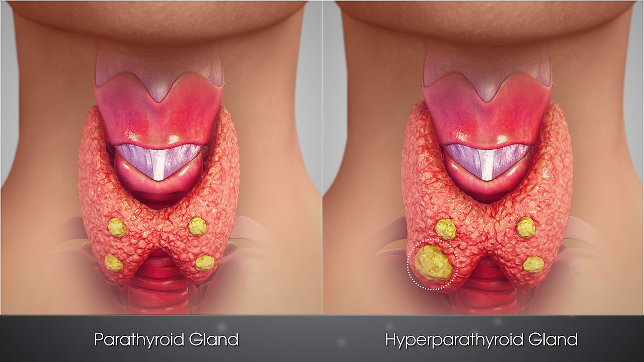 Parathyroid glands (from Wikipedia)
I thought it may be best to illustrate a typical journey of a patient with primary hyperparathyroidism by describing a patient I saw late last year. The patient saw me in our ENT Clinic at Mt Elizabeth Novena Hospital.
A case study
FT is a bubbly and active 58-year-old lady who presented to me in October 2020. She had a long history of high blood calcium that was found on routine blood tests but often dismissed by her primary care physician. In fact, to help prevent osteoporosis, she was once given calcium supplements! In August 2020, her calcium level had reached 2.78 (normal range up to 2.60 mmol/L). A sharp family physician noticed this and did a special assay for her parathyroid hormone (PTH) level. This was raised at 9.84 (normal range up to 9.60 pmol/L) and her vitamin D levels were normal.
She was promptly sent to the specialists in a local hospital who ordered an ultrasound scan and a special investigation called a SestaMIBI scan. The SestaMIBI scan is a nuclear medicine scan. These scans showed a parathyroid adenoma but the results were discordant. The ultrasound suggested a left-sided adenoma but the nuclear medicine scan showed a right-sided adenoma.
When I saw her, I repeated the scan in my office. I saw a small dark nodule behind the right thyroid lobe
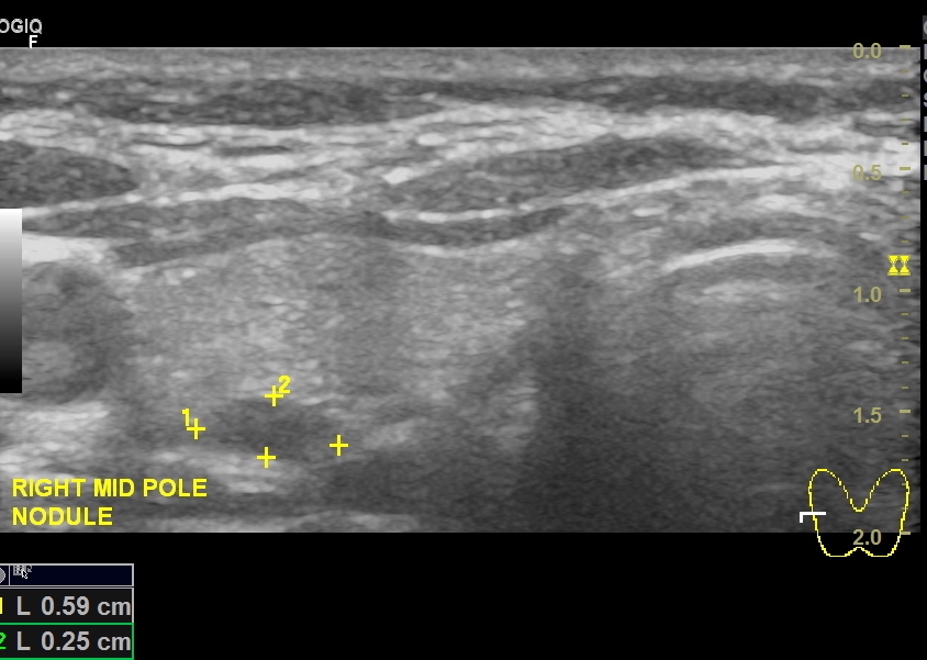 Ultrasound scan showing a dark (hypoechoic) nodule behind the right thyroid lobe
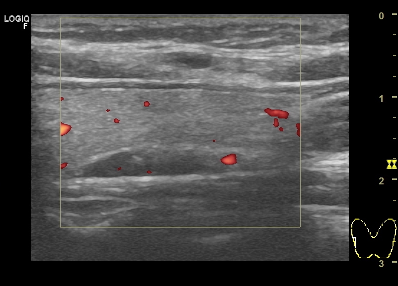 “Polar vessel” sign – suggestive of a parathyroid nodule
I thus ordered a SestaMIBI scan. This nicely demonstrated a right-sided adenoma. We had located the culprit!
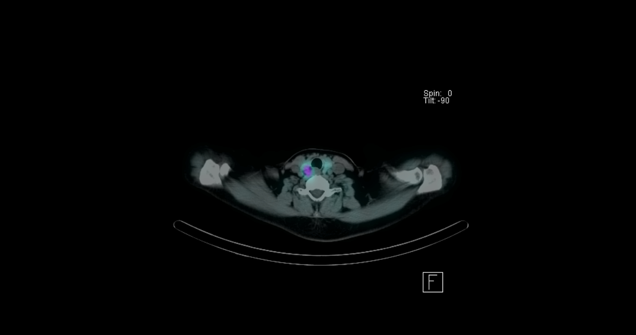 SestaMIBI scan showing a right parathyroid adenoma
A few weeks later, FT was taken to the operating theatre. She was placed under general anaesthesia and I infused a saline solution mixed with methylene blue through her veins. Methylene blue has a propensity to stain parathyroid tissue and it makes their identification easy.
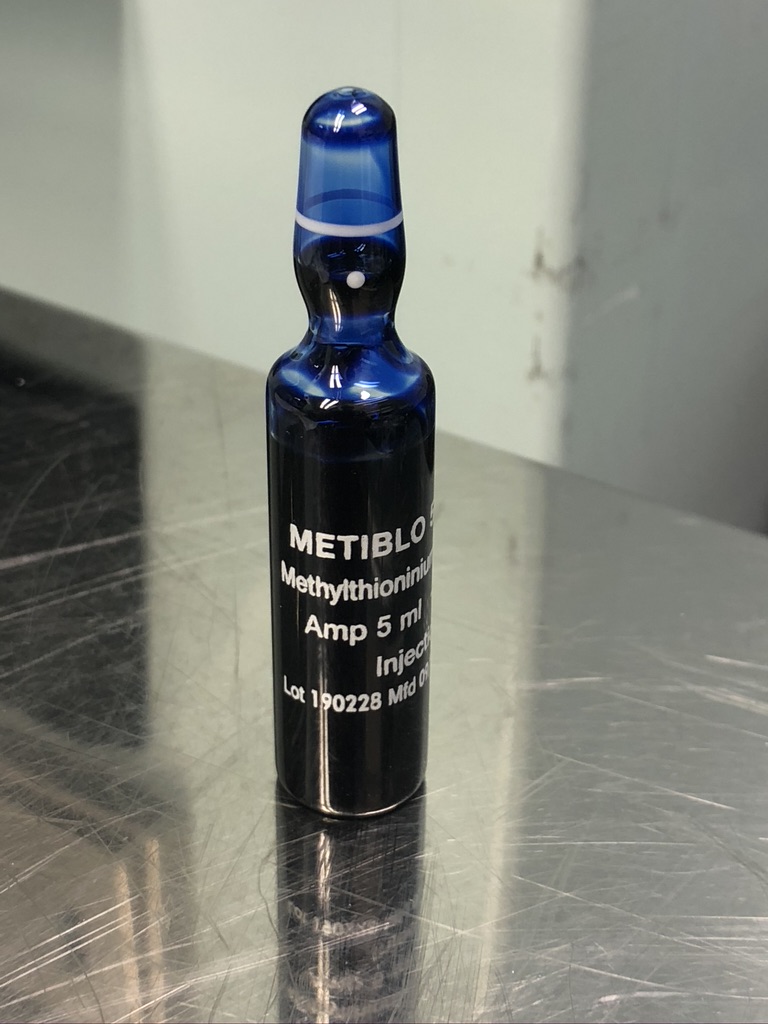 Methylene Blue
Under good magnification, the parathyroid adenoma was identified and confirmed. I also removed a portion of a second parathyroid gland to establish that it was normal.
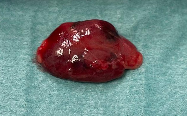 Parathyroid adenoma
FT made a splendid recovery. Her calcium levels improved and she felt so much better for it!
Share this blog via:


