


Chronic sinusitis – imaging your sinuses
Sinusitis is the inflammation of the paranasal sinuses. The paranasal sinuses are bony cavities within the skull that communicate with the nasal cavity. The passage from these sinuses to the nose is often narrow and any inflammation of the lining of these passages can cause obstruction. When sinusitis is prolonged and lasts more than 12 weeks, we use the term chronic sinusitis. Chronic does not mean severe. It simply means that condition has not improved over time.
The diagnosis of sinusitis is made by examination. It usually involves nasal endoscopy which is the fibreoptic endoscopic examination of the nasal cavity. When nasal endoscopy is not readily available, imaging of the sinuses may be helpful. The simple plain x-ray of the sinuses was a common investigation. This x-ray of the face has little utility in modern practice. Perhaps its only use is in young children who cannot tolerate nasal endoscopy.
The most useful form of imaging of the sinuses is the CT scan (or computed tomography). The CT scan of the sinuses is a quick investigation lasting no more than 15 minutes. It uses x-rays and advanced computer technology to reconstruct a detailed image of your nose and sinuses. For this examination, you will be required to remove spectacles, hearing aids, ear rings, necklaces and any false teeth. These items can interfere with this form of imaging. You will then be asked to lie flat on the bed that can be moved slowly into the scanner.
A patient having a CT scan of the sinuses. Image from ENT Today
The standard CT scan of the sinuses for chronic sinusitis that ENT surgeons request does not involve an injection of a contrast medium or dye into your veins. If your ENT surgeon is requesting a scan because she is concerned about a tumour in the nose or sinuses, she may request the radiographer to insert an intravenous cannula into your hand and inject a contrast medium whilst you have the scan. This is called a contrast-enhanced CT scan of the sinuses.
How much radiation does a CT sinus deliver?
This is a common question that many patients ask. The natural concern is that the radiation received is harmful and may cause cancer. The amount of radiation that a person receives is measured in millisieverts (mSv). This is often described as the effective dose. In most countries the background environmental radiation delivers an effective dose of 3 mSv per annum. A standard CT sinus scan delivers approximately 2 mSv of radiation. This is equivalent to 8 months of radiation delivered in 15 minutes! The USFDA has an advisory on the risk of cancer from having a CT scan. It is estimated that a CT examination that delivers an effective dose of 10 mSv increases the risk of a fatal cancer by 1 in 2000 over one’s lifetime. So the increased risk from a CT scan of the sinuses (2 mSv) is very small but not negligible. ENT surgeons should only perform scans when it may alter a patient’s management. The nasal endoscope should remain the primary method of diagnosing and monitoring chronic sinusitis.
A normal CT sinus
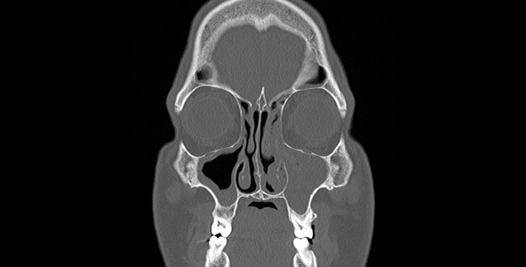
The CT sinus images below are of a patient with headaches which were thought to be sinus in origin. The nasal endoscopy was normal but the symptoms were highly suggestive of sinusitis. A CT sinus was obtained and these images show completely normal paranasal sinuses.
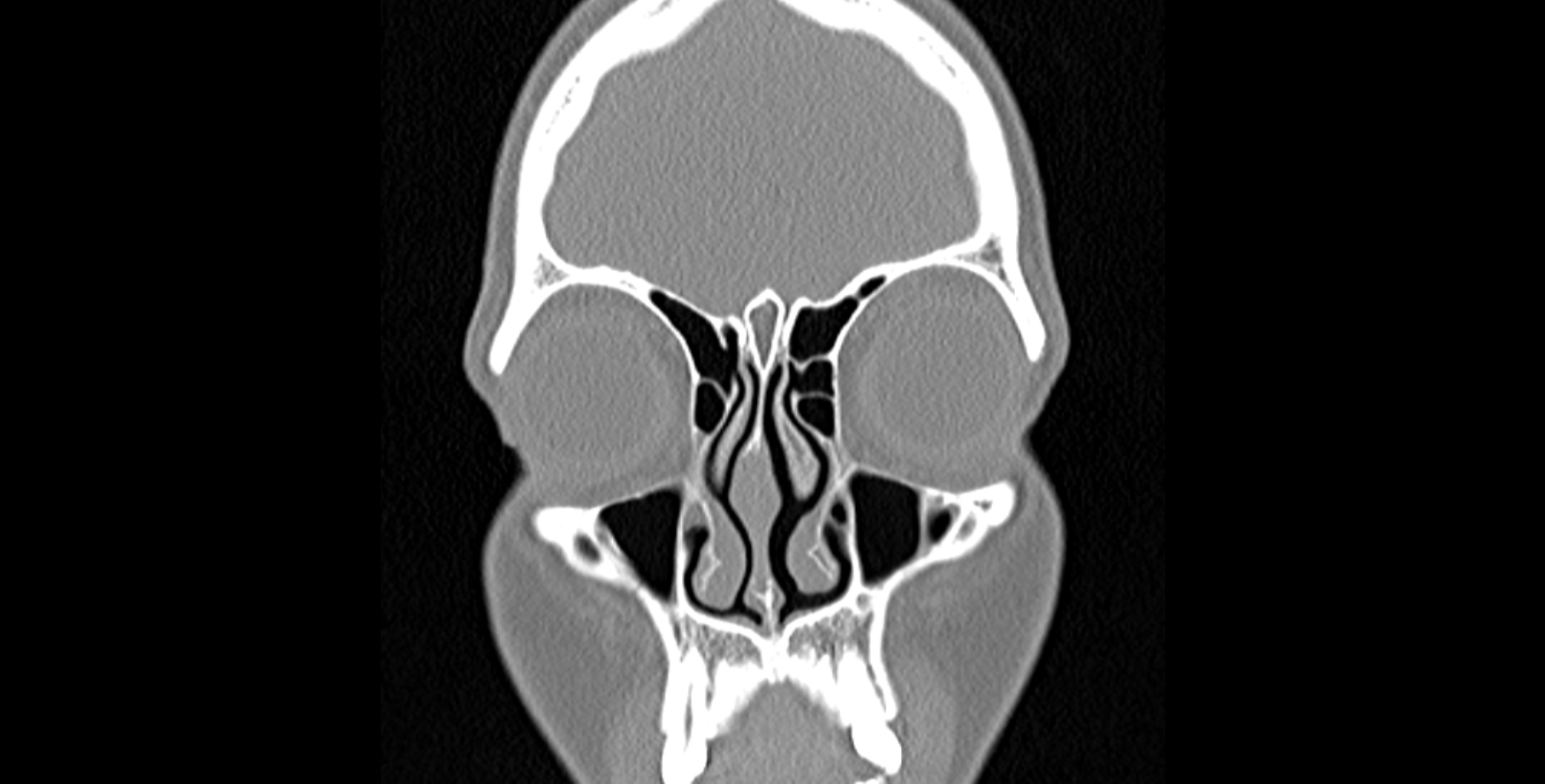
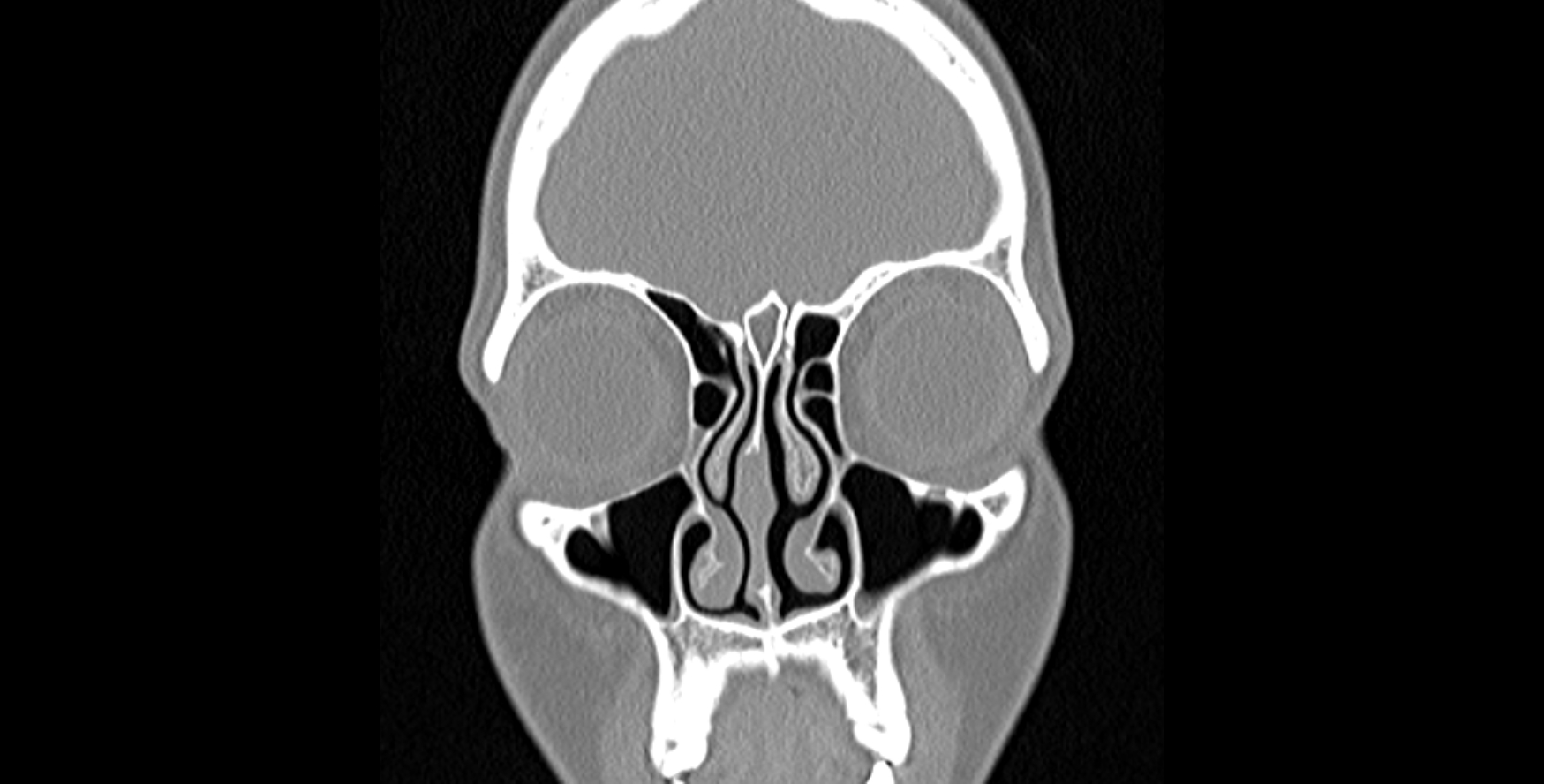
These scans are easy to read. All you need to know is that bone and bony structures appear white, air appears black and soft tissue or fluid appear grey. These scans are called coronal sequences or slices. It is as if the patient is facing you through a window! Structures on the right are on the patient’s left. The eyes are easily made out. The cheek sinuses known as the maxillary sinuses appear as black spaces below the eyes.
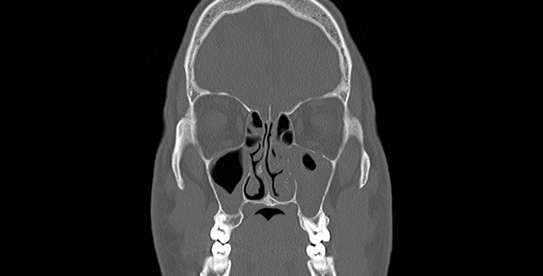
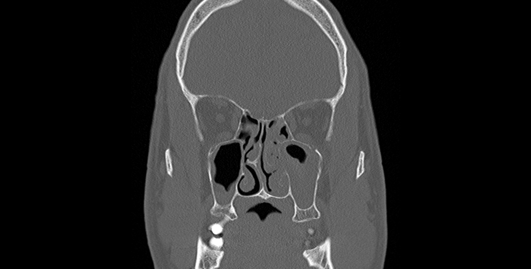
CT sinus showing chronic sinusitis
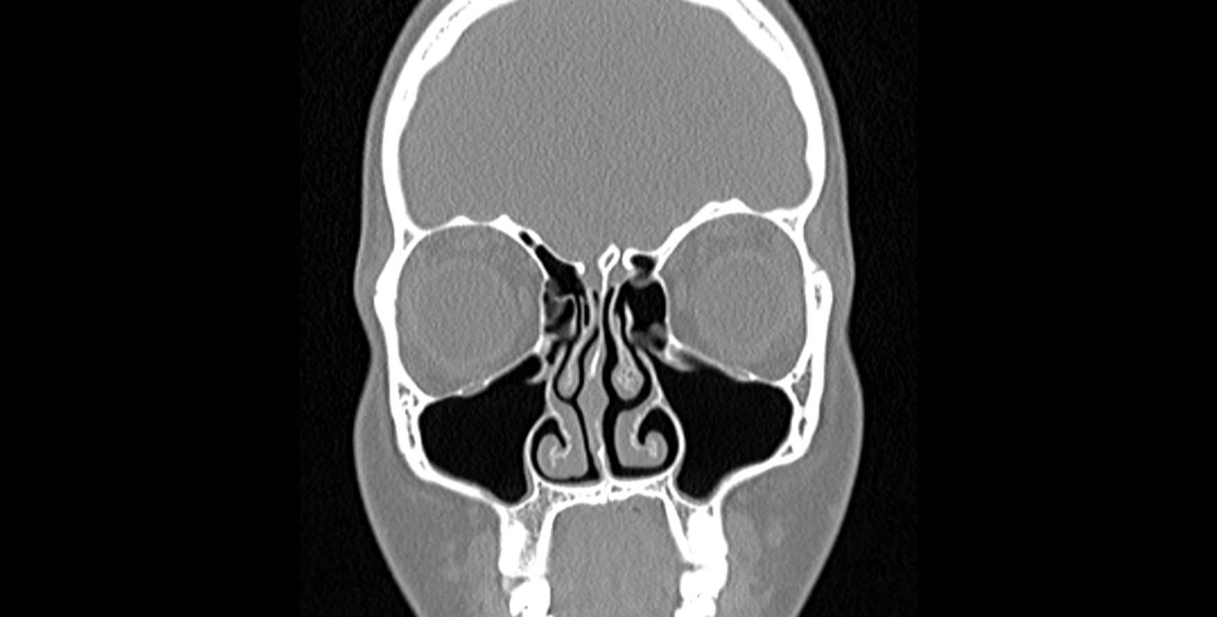
The scans below show chronic sinusitis. As you can tell the air-filled sinus spaces are not completely black. They are opacified – suggesting the sinus lining is thickened or there is fluid or pus in the sinuses. In this patient you may also see that the wall of bone and cartilage that separated the nose – right from left – is not straight. This structure is called the septum and it is deviated in this patient with a prominent spur to the patient’s right.
This patient failed to repond to medical treatment and went on to have surgery. Surgery for chronic sinusitis is called Functional Endoscopic Sinus Surgery and I will come to talk about this in another blog soon!
END
Share this blog via:


